Treating Compression Fracture with Kyphoplasty vs. Bracing

Vertebral Compression Fractures: Bracing vs Outpatient Kyphoplasty

written by: Dr. Thuy M. Nguyen

What is a compression fracture?

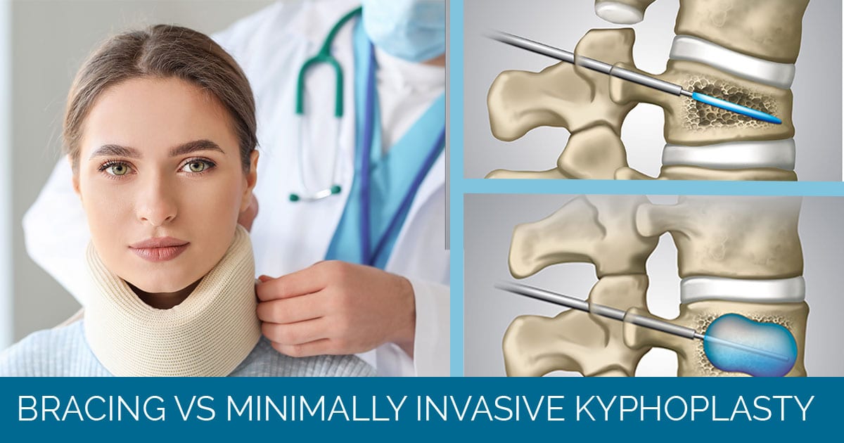
A compression fracture is a fracture involving the front third of your vertebral body. This will commonly have an appearance of wedging on X-ray. Compression fractures can be very painful with associated point tenderness.

Bracing Compression Fractures

Kyphoplasty is a minimally invasive spine procedure for treating compression fractures. As compression fractures are usually stable, I typically like to treat these with bracing and analgesics. We will do a trial of conservative management as surgery should be a last resort. Bracing is employed to decrease motion at the fracture which is responsible for inducing pain. A back brace also serves to reduce repetitive “microfractures”. If the back brace and conservative management is not reducing the amount of pain to satisfaction, then a discussion regarding a kyphoplasty should be undertaken.

What is Kyphoplasty?

Kyphoplasty is a minimally invasive procedure that I do in the office with a local numbing medication. No general anesthesia is used to ensure quicker recovery and a same day procedure as well as avoids all the risk factors and side effects that accompany general anesthesia. My patients will come into the office for the procedure and leave after the procedure.  The kyphoplasty is done under xray or fluoroscopy in order to guide the needle into the correct location. A cavity is created within the vertebral body and a synthetic medical grade “cement” is introduced. This “cement” is polymethlymethacrylate or PMMA.  The PMMA will mend the fractured pieces together and therefore stop motion at these fracture segments which is responsible for the pain.

About Dr. Thuy M. Nguyen

Dr. Nguyen received his training in neurosurgery in a highly sought after residency program at Beaumont Health in Detroit. He then pursued a fellowship in orthopaedic spine surgery at the Oregon Health and Science University. He brings a unique set of spine-focused clincal acumen to Joseph Spine Institute.

About Joseph Spine Institute

Joseph Spine Institute is the advanced center for spine, scoliosis and minimally invasive spine surgery. Founded by Dr. Samuel A. Joseph, Jr., a fellowship trained, board-certified orthopedic surgeon. Dr. Joseph has assembled a comprehensive team of highly skilled spine specialists that are devoted to offering the highest level of spine care available today. He has also strategically partnered with some of the most innovative leaders in the spine care industry today in order to bring our patients the latest in technology, products and resources.

From state-of-the-art technology, to alternate therapies and interventional pain management treatments our industry leading spine care practice offers our patients a comprehensive, multidisciplinary approach to diagnosis, treatment and rehabilitation of spinal conditions. Our highly-trained surgeons offer an integrated approach to patients that includes surgical and non-surgical options such as pain management and physical therapy.

minimally invasive spine surgery tampa bay

The Difference Between Minimally Invasive Spine Surgery vs. Traditional Open Surgery

The Difference Between Minimally Invasive Spine Surgery vs. Traditional Open Surgery

written by Dr. Samuel A. Joseph, Jr.

Spine surgery has been transformed significantly by the growth of minimally invasive spine surgery (MISS) procedures. As a spine surgeon I am always researching new ways to improve patient outcomes. Implementing new advancements that improve recovery time, create less scaring and less pain after an operation is the ultimate goal for all of us at Joseph Spine Institute. By combining advanced techniques and partnering with with innovators developing cutting edge spine technologies patients now have more options for minimally invasive spine surgery and outpatient procedures than ever before.

Is Minimally Invasive Spine Surgery Right for You?

Many patients come in to my office confused about the difference between open surgery and minimally invasive spine surgery otherwise called MIS. The traditional open surgery is still necessary in certain situations. However, with the advancement of newer technology most procedures can now be performed in a MIS manner. There are many factors involved in this decision, most importantly are the specific details of your condition. Consulting an exceptionally trained and well respected board certified spine surgeon is of the utmost importance when determining the type of treatment options that are best for you.

What Is Open Spine Surgery?

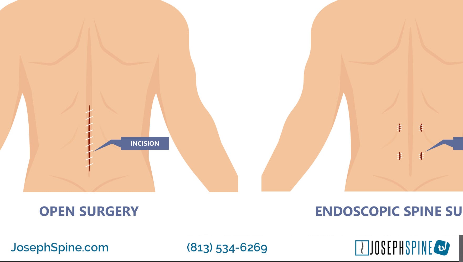
During open surgery, you can expect to be opened up (hence, the name). Open surgery involves making an incision that then proceeds down to pull the muscles and ligaments off the spine to get access to the problem area. This is typically done in a hospital and requires an overnight stay. It’s also possible that, during an open procedure, muscles and tendons will have to be cut through and moved aside to reach the affected area. This can cause damage to these tissues, possible large loss of blood and prolong recovery.

Once bones of the spine can be visualized, the necessary spinal procedure can begin. Most open spine surgeries require the following:

  • Anesthesia
  • Large incisions
  • Muscle retraction
  • Long surgery times
  • Hospitalization
  • Long recovery times

What is Difference with Minimally Invasive Spine Surgery?

MIS surgery approaches the same problem, but with a different rationale. The goal is to preserve the muscles and tissues through smaller incision and techniques that go through or around the muscles without stripping them off.

MIS is performed using much smaller incisions than open surgery. Small tube shaped retractors, rather than large muscle dissecting ones, are used to create a narrow opening to accesses the spine.  X-ray fluoroscopy and a microscope allow me to visualize the area of interest in the spine.

Very small instruments are used to remove pieces of damaged vertebral bone, cartilage, and/or spinal disc. This type of minimally invasive approach results in less damage to the muscles and soft tissues that surround the spine, which leads to an expedited recovery and less post-operative pain.

What are the benefits of minimally invasive spine surgery?

  • Less OR time for procedure
  • Faster recovery
  • Less bleeding (which means little chance of blood transfusion)
  • Smaller incision
  • Little or no muscle cutting
  • Less risk of infection
  • Less pain after surgery
    • Decreased reliance on pain medication
  • Less rehabilitation is needed
    • Patients return to work and activities more quickly
  • Less scarring

“MIS surgeries are usually done outpatient which means no hospital stay and getting home that day.  This allows me as a spine surgeon to get you back to life much faster!” Dr. Samuel A. Joseph.

Minimally Invasive Surgical Procedures

The following spinal surgeries can be performed using a minimally invasive technique:

  • Spinal Fusion – Spinal fusion is surgery to permanently connect two or more vertebrae in your spine, eliminating motion between them. Spinal fusion involves techniques designed to mimic the normal healing process of broken bones. During spinal fusion, your surgeon places bone or a bonelike material within the space between two spinal vertebrae. Metal plates, screws and rods may be used to hold the vertebrae together, so they can heal into one solid unit.
  • Kyphoplasty – Kyphoplasty is a surgical procedure that expands and stabilizes compression fractures of the spine. Kyphoplasty uses a small balloon that gently lifts bone fragments into their correct position.  The balloon is inflated to create a space that the surgeon later fills with a special bone cement. The bone cement creates an internal cast that holds the vertebra in place. This extra feature allows kyphoplasty to help restore vertebral height, and thus, help reduce spinal deformity.  It also allows a thicker cement to be used which has less risk of leaking out and causing complications.
  • Cervical Disc Replacement – Cervical disk replacement surgery involves removing a diseased cervical disk and replacing it with an artificial, man-made device. An artificial disc may be used to replace a disc in either the lumbar spine (lower back) or cervical spine (neck).
  • Laminectomy – A laminectomy is a procedure to remove the spinous process and lamina of the affected vertebra. The surgery relieves pressure on the spinal nerve roots by creating more space in the spinal canal. This procedure is primarily performed to relieve symptoms of spinal stenosis.
  • Discectomy and Microdiscectomy – Removes either a part of or an entire intervertebral disc to relieve nerve pressure. The difference between the two procedures is that a microdiscectomy is performed using an operating microscope. These procedures are used to remove either ruptured or herniated discs.
  • Spinal Cord Stimulation – A pain management technique that involves the surgical implantation of an electrotherapeutic device onto the spinal cord. In the procedure, a device is implanted that introduces low levels of electrical current to the dorsal portion of the spinal cord to block the sensation of pain. Spinal cord stimulators may be a fully implanted system or a system with an external power source.
  • Interspinous Spacer Placement A spacer is an implant that sits between two interspinous processes in the lumbar spine. These spacers are inserted in a minimally invasive procedure. The purpose of interspinous spacers is to increase the space between processes and relieve the symptoms of spinal stenosis

What spinal conditions can be treated with MIS?

  • Herniated Disc – A condition in which the annulus fibrosus (outer portion) of the vertebral disc is torn, enabling the nucleus (inner portion) to herniate or extrude through the fibers. The herniated material can compress the nerves around the disc and create pain that can radiate through the back and sometimes down the arms (if the herniation is in the cervical spine) and legs (if the herniation is in the lumbar spine).
  • Degenerative Disc Disease (DDD) –  A condition in which a damaged spinal disc causes pain. (While everyone has some wear and tear of their spinal discs; not everyone will have pain.) And although it may take decades before you feel pain (if you ever do), the degeneration process can start as early as your 20s.
  • Sciatica – Sciatica refers to pain that radiates along the path of the sciatic nerve, which branches from your lower back through your hips and buttocks and down each leg. Typically, sciatica affects only one side of your body. Sciatica most commonly occurs when a herniated disk, bone spur on the spine or narrowing of the spine (spinal stenosis) compresses part of the nerve. This causes inflammation, pain and often some numbness in the affected leg.
  • Scoliosis, Spinal Deformities – Scoliosis and other types of spinal deformity are characterized by abnormal spinal curves that can vary in size and shape. Normal curvature of spine, called lordosis and kyphosis, naturally occur at different levels or regions of the spinal column. However, scoliosis and/or excessive lordotic or kyphotic curves (eg, hyperkyphosis) are abnormal types of spinal curvatures that can affect balance, flexibility and spinal alignment.
  • Spinal Stenosis – A narrowing of the spaces within your spine, which can put pressure on the nerves that travel through the spine. Spinal stenosis occurs most often in the lower back and the neck.
  • Vertebral Compression Fracture (VCF) – occur when the bony block or vertebral body in the spine collapses, which can lead to severe pain, deformity and loss of height. These fractures more commonly occur in the thoracic spine (the middle portion of the spine), especially in the lower part.
  • Spondylolisthesis – A spinal disorder in which a bone (vertebra) slips forward onto the bone below it.
  • Spinal Infections – An infectious disease that affects the vertebral body, the intervertebral disk, or adjacent paraspinal tissue. Spinal infections can develop after a surgical procedure or arise on their own. Infections can affect different parts of your spine—for example, some infections develop in a vertebral body while others may affect an intervertebral disc. Constant back pain without prior injury is a red flag symptom of spinal infection.
  • Spinal Tumors –  A growth that develops within your spinal canal or within the bones of your spine. A spinal cord tumor, also called an intradural tumor, is a spinal tumor that that begins within the spinal cord or the covering of the spinal cord (dura). A tumor that affects the bones of the spine (vertebrae) is called a vertebral tumor.

Due to the rapid advance in technology new techniques, procedures, equipment and implants are developed to further enhance the treatment of spine disorders and diseases through minimally invasive procedures. However, it is important to note that some severe cases of spinal infections, scoliotic curves, or complex multi-level spinal procedures cannot be treated minimally invasively.

About Dr. Samuel Joseph

Dr. Samuel Joseph is a board certified, fellowship-trained, orthopaedic surgeon who specializes in advanced spine care. He is a leader in new spine medicine techniques and procedures.  He is dedicated to pioneering minimally invasive, outpatient spine surgical techniques along with complex scoliosis and revision surgery. Dr. Joseph is among a select group of surgeons to offer cutting edge endoscopic and robotic-assisted surgery for patients to provide the most advanced minimally invasive spine surgery (MIS) available globally. He is also the team spine surgeon for the Tampa Bay Buccaneers.

About Joseph Spine Institute

Joseph Spine Institute is the advanced center for spine, scoliosis and minimally invasive spine surgery. Founded by Dr. Samuel A. Joseph, Jr., a fellowship trained, board-certified orthopedic surgeon. Dr. Joseph has assembled a comprehensive team of highly skilled spine specialists that are devoted to offering the highest level of spine care available today. He has also strategically partnered with some of the most innovative leaders in the spine care industry today in order to bring our patients the latest in technology, products and resources.

From state-of-the-art technology, to alternate therapies and interventional pain management treatments our industry leading spine care practice offers our patients a comprehensive, multidisciplinary approach to diagnosis, treatment and rehabilitation of spinal conditions. Our highly-trained surgeons offer an integrated approach to patients that includes surgical and non-surgical options such as pain management and physical therapy. 

endoscopic fusion spine minimally invasive

New Endoscopic Spine procedure developed by Dr. Sam Joseph

28 Apr 2021 Spine News

Endoscopic Spine Fusion New Minimally Invasive Approach Developed by Dr. Joseph

In this post I will explain an innovative new minimally invasive technique that I have personally developed in an ongoing effort to offer my patients the very best surgical methods available.

This innovative new Transforaminal lumbar endoscopic interbody fusion or an (ETDIF) is a revolutionary minimally invasive spinal fusion technique that combines the precision of the endoscope and a new break through on spinal fusion implant that features an expandable cage.

An endoscopic ETDIF is similar to an open ETDIF surgery, but includes the use of a minimally invasive approach with a tiny camera (the endoscope) that sends images to a monitor. This technique may eliminate the need for anesthesia with less risk and faster recovery.

An endoscopic approach involves tube(s) inserted into the operative area using relatively small incisions, with a camera to provide visibility to the surgeon and small instruments inserted through the tube(s) to conduct the operation.

Confining all instruments to the tube (or sometimes two tubes) is designed to minimize disruption to surrounding tissue and bone, with the goal of less tissue damage and faster healing times.

Preparation for the Endoscopic Spine Procedure

• The patient lies face down to prepare for the procedure. General anesthesia may be used. There is a good possibility that general anesthesia may be avoided in select patients.

• An incision of about one centimeter is made on the most symptomatic side of the spine at the level of the problematic disc.

• Fluoroscopic (live X-ray) visualization is used to identify the appropriate disc.

Preparing the disc space for fusion

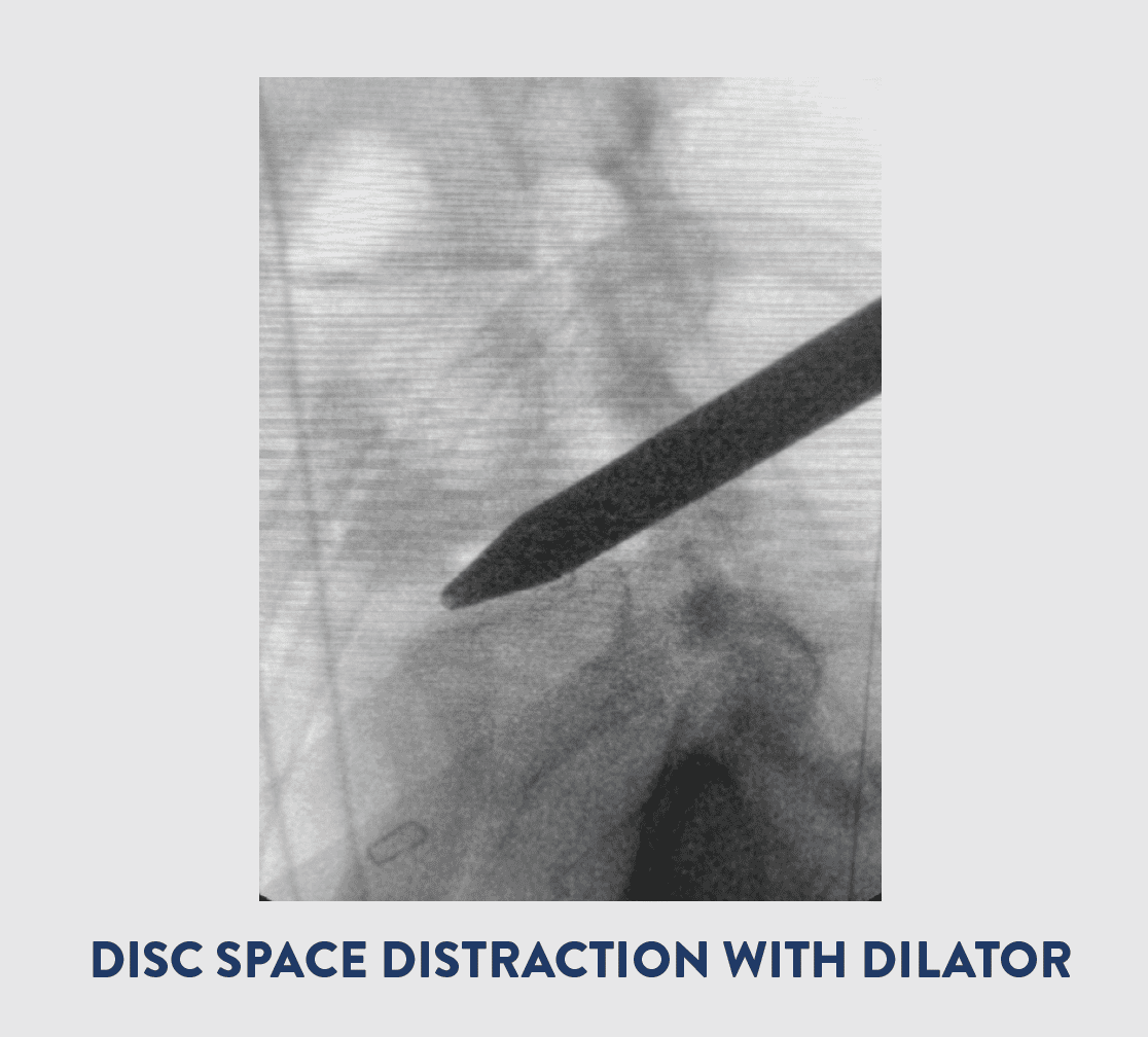
• A thin tube is inserted through the incision and into the disc area through the neuroforamen opening, the channel through which the nerves exit the spinal canal. A dilator is used to enlarge the size of the tube and provide a working channel for the operative instruments.

• The endoscope is inserted through the tube into the disc to allow the surgeon to assess the disc.

• The disc area is irrigated with antibiotic solution minimizing the risk of post-operative infection.

• Special instruments are inserted into the tube(s) to remove damaged portion of the disc, and the parts that are cut away are extracted through the tube(s).

• Cartilage covering the surfaces of the bones bordering the disc is removed and extracted through the tube.

• Dilators are then inserted into the disc to restore the disc height and normal spacing between the vertebrae, as necessary.

• This concludes the part of the operation that prepares the disc space for the fusion. The success of the disc preparation is confirmed endoscopically. This part of the process is essential to ensure that bone graft inserted into the disc space is likely to fuse to the bone surfaces bordering the disc.

Inserting Expandable Cage Spinal Implant

• The cage is inserted into the disc space. The cage provides a minimal insertion profile, designed with less neural retraction.

• This expansion allows for maximum graft delivery. The volume is only restricted by amount of disc that is removed.

• Absorbable sutures are used to close the wound and the surgical track is infiltrated with local anesthetic for immediate post-operative pain control. Antibiotic dressing is placed on the wound.

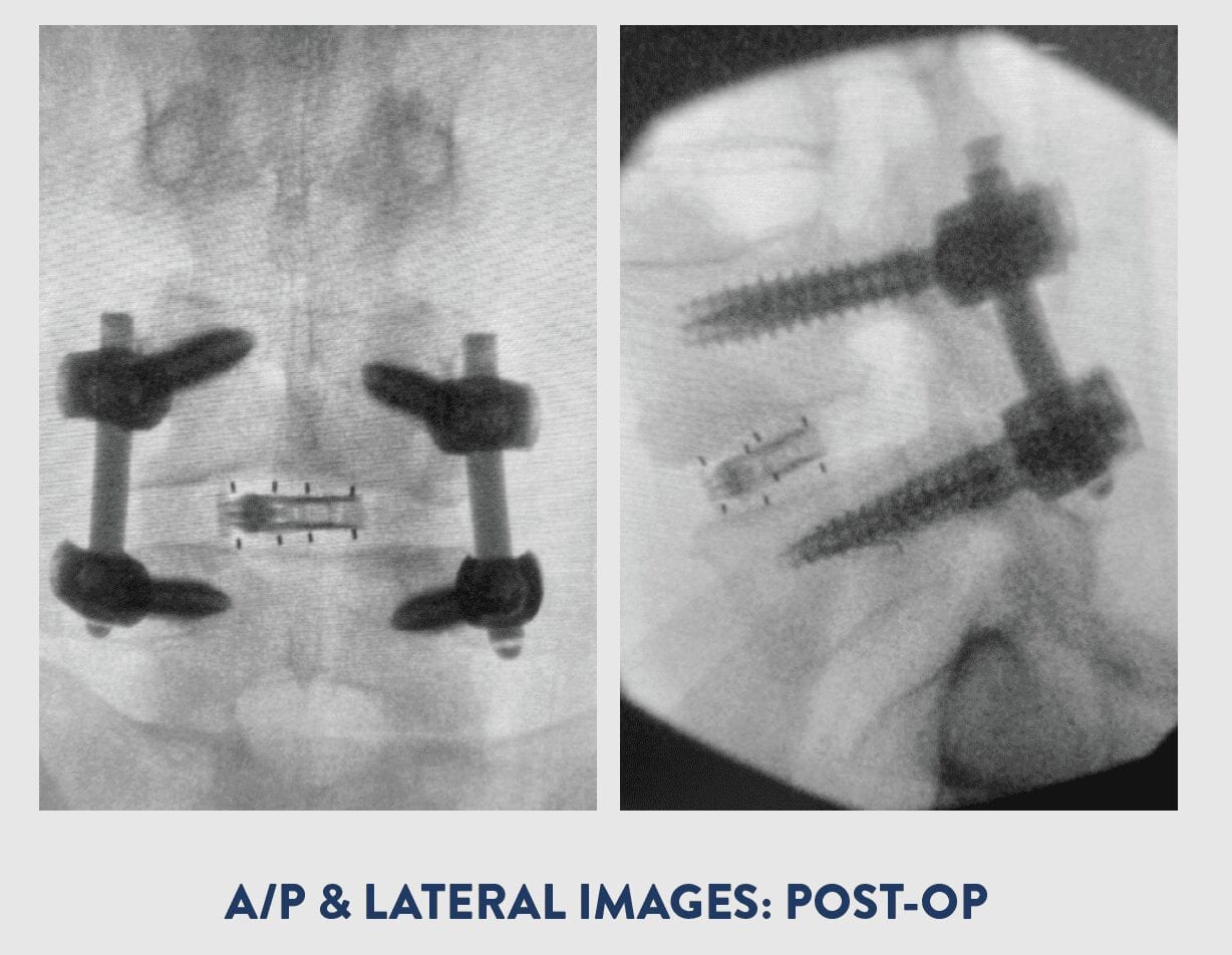
In some cases, screws, reinforced with rods, may be attached to the back of the vertebra to add stability and prevent movement in the area while the bone graft sets. The bone graft fosters bone growth around these rods, and they are typically left in place even after the bone graft takes hold.

The procedure is approximately 1 1/2 to 2 hours and is usually performed as an outpatient surgery.

About Dr. Joseph
Dr. Joseph is a board certified, fellowship-trained, orthopaedic surgeon who specializes in advanced spine care. He is a leader in new spine medicine techniques and procedures. He is dedicated to pioneering minimally invasive, outpatient spine surgical techniques along with complex scoliosis and revision surgery. Dr. Joseph is among a select group of surgeons to offer cutting edge endoscopic and robotic-assisted surgery for patients to provide the most advanced minimally invasive spine surgery (MIS) available globally. He is also the team spine surgeon for the Tampa Bay Buccaneers.

About Joseph Spine Institute

Joseph Spine Institute is the advanced center for spine, scoliosis and minimally invasive spine surgery. Founded by Dr. Samuel A. Joseph, Jr., a fellowship trained, board-certified orthopedic surgeon. Dr. Joseph has assembled a comprehensive team of highly skilled spine specialists that are devoted to offering the highest level of spine care available today. He has also strategically partnered with some of the most innovative leaders in the spine care industry today in order to bring our patients the latest in technology, products and resources.

From state-of-the-art technology, to alternate therapies and interventional pain management treatments our industry leading spine care practice offers our patients a comprehensive, multidisciplinary approach to diagnosis, treatment and rehabilitation of spinal conditions. Our highly-trained surgeons offer an integrated approach to patients that includes surgical and non-surgical options such as pain management and physical therapy. 

Dr. Samuel A. Joseph Jr, First in the Nation to Perform Outpatient Prone Lateral Fusion

Dr. Samuel A. Joseph Jr, First in the Nation to Perform Outpatient PTP (Prone Transpsoas)

Physicians like Dr. Samuel Joseph, founder of Joseph Spine Institute in Tampa Florida, are constantly researching and developing new ways to improve minimally invasive spine techniques for their patients. Dr. Joseph works in conjunction with other leaders in spine research, development and technology to bring minimally invasive procedures to complex spine conditions, which in turn results in better faster recovery for patients.

Take for example a traditional lateral interbody fusion (LIF) that has a patient lying in a lateral position. Not only can the position be unfamiliar to the surgeon and staff which leads to inefficiencies in preoperative setup and repositioning for posterior procedures, but literature demonstrates that the lateral decubitus position may not be ideal for achieving sagittal alignment – the greatest correlative to a long-term positive outcomes.

The newly developed procedure performed by Dr. Joseph, PTP (Prone Transpsoas) has many benefits that range from superior surgical efficiency to powerful correction abilities in both the sagittal and coronal planes.  PTP allows for the patient to remain in one position for the entire procedure. The prone position creates efficiencies by allowing for a single-position surgery providing simultaneous access to the anterior column and posterior column of the spine. 

View Published Case Study

Initial multi-center clinical experience with prone transpsoas lateral interbody fusion: Feasibility, preoperative outcomes, and lessons learned.

About Dr. Joseph

Dr. Joseph is a board certified, fellowship-trained, orthopaedic surgeon who specializes in advanced spine care. He is a leader in new spine medicine techniques and procedures.  He is dedicated to pioneering minimally invasive, outpatient spine surgical techniques along with complex scoliosis and revision surgery. Dr. Joseph is among a select group of surgeons to offer cutting edge endoscopic and robotic-assisted surgery for patients to provide the most advanced minimally invasive spine surgery (MIS) available globally. He is also the team spine surgeon for the Tampa Bay Buccaneers.

About Joseph Spine Institute

Joseph Spine Institute is the advanced center for spine, scoliosis and minimally invasive spine surgery. Treatment includes therapeutic injections, physical therapy, pain management, stem cell therapy, disc replacement surgery, minimally invasive techniques, including robotic-assisted surgery, and complex reconstruction of adult and pediatric spinal disorders. Dr. Joseph is among a select group of surgeons to offer cutting edge endoscopic and robotic-assisted surgery for patients to provide the most advanced minimally invasive spine surgery (MIS) available globally. Joseph Spine has four locations in Tampa Bay, including Tampa, St. Petersburg, Safety Harbor, and Brandon, FL. Joseph Spine is dedicated to offering humanitarian spine services internationally in the form of education, awareness, and surgery. Please visit JosephSpine.com for more information.

Dr. Joseph Performs Endoscopic Decompression and Fusion with Coronal Correction

Dr. Sam Joseph continues to pioneer new techniques for minimally invasive spine surgery by implementing cutting edge techniques and utilizing the FlareHawk7 endoscopically assisted system.

CLINICAL PRESENTATION

The patient is a 52-year-old female with a longstanding history of severe lower back and right leg pain. She had failed therapy, medication and injections, and had a prior right-sided laminectomy at L4-5. The patient presented to another surgeon who offered her open lumbar laminectomy and fusion. She then presented to Dr. Joseph’s office for a second opinion for a less invasive approach.

EXAM

Severe lower back pain with palpation, spasm, pos SLR right side, decreased sensation L3, 4, 5, absent DTR right side.

IMAGING

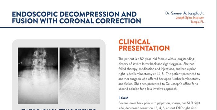
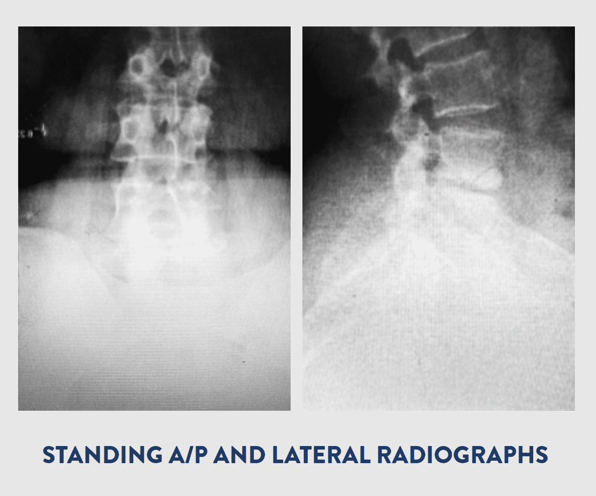
X-RAY

  • Moderate spondylitic changes at L4-5
  • Endplate degeneration identified
  • Facet arthropathy identified
  • Scoliosis curvature
  • Exhale. Leave the pose incrementally. First, bend the arms slowly backwards. Lower the navel to the mat, then the chest, then the shoulders, and, lastly, the forehead.

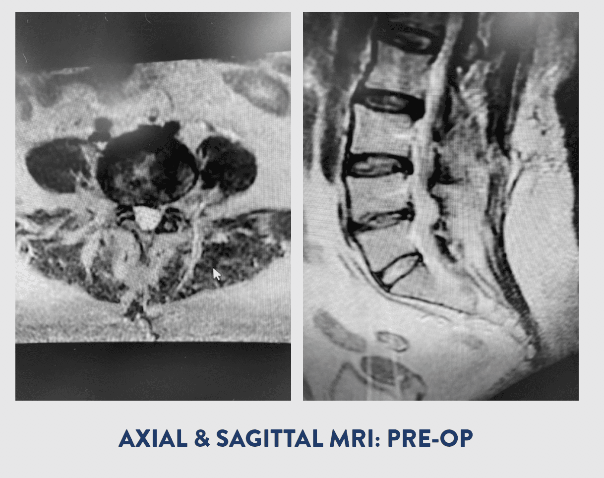
MRI LUMBAR SPINE

  • L2-3: 5 mm central disk protrusion is seen with bilateral facet arthropathy.
  • L3-4: Dehydrated, bulging disc is seen with a 4 mm broad-based disc protrusion and bilateral facet arthropathy.
  • L4-5: Dessicated, bulging disc and osteophyte and facet arthropathy are seen with a 7 mm right foraminal disc protrusion. Prior Laminectomy. Severe foraminal narrowing on the right side.
  • Exhale. Leave the pose incrementally. First, bend the arms slowly backwards. Lower the navel to the mat, then the chest, then the shoulders, and, lastly, the forehead.

Modified Seated Spinal Twist

Modified Seated Spinal Twist

PROCEDURE

OVERVIEW

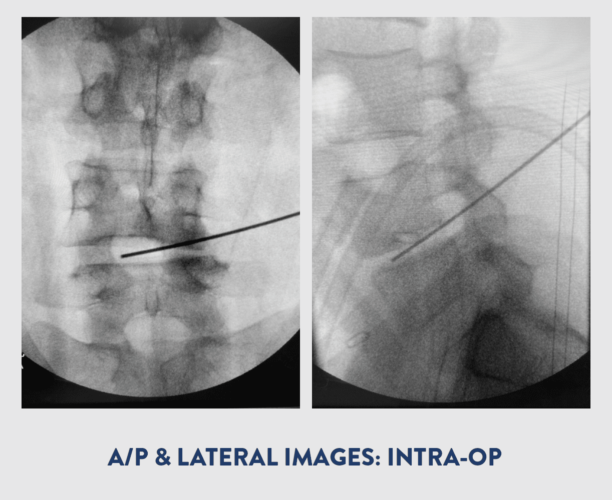
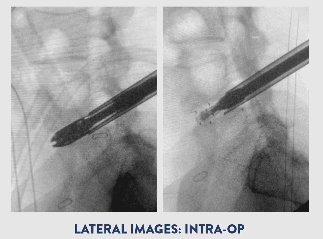
Dr. Joseph performed a right sided foraminotomy, discectomy, and partial facetectomy of L2-3, L3-4 and L4-5. An endoscopically assisted TLIF was performed on the right side of L4-5 with bilateral posterior pedicle screw instrumentation at L4-5.The FlareHawk7 endoscopically assisted system allowed Dr. Joseph to perform his discectomy and deploy an 11mm wide, 26mm long, 10mm tall Implant with 6° of lordosis through an 11mm Tubular Retractor.

BLOOD LOSS

50 cc

IMPLANT INSERTION PROFILE

7mm W x 7mm H x 26mm L

IMPLANT DEPLOYED PROFILE 11mm W x 10mm H x 26mm L with 6° Lordosis

PROCEDURAL RATIONALE

Endoscopic decompression with fusion was ideal for this patient due to her multilevel symptomatic foraminal stenosis as well as her scoliosis and prior Midline Laminectomy at L4-5. The Kambin’s triangle approach avoids the scar tissue from prior surgery. The round dilator acts like a distractor elevating the right side and the FlareHawk7 allows expansion to maintain the correction through a very small working channel. This allows both direct and indirect decompression at the L4-5 level.

OUTCOME

The patient had no more leg pain and only mild back soreness. Discharged home in fewer than 24 hours post-op. The patient continues to do well.

Voted Top Spine Surgeon Tampa Bay – Dr. Samuel A. Joseph, Jr.

18 Aug 2020 Spine News

Voted Top Spine Surgeon for Tampa Bay

Dr. Samuel A. Joseph Jr., a leader in minimally invasive spine surgery, and founder of Joseph Spine Institute, was voted by his peers to Tampa Magazines’ 2020 Top Doctors list in the specialty of Orthopedic Spine Surgery for the third straight year in a row.

In May, Tampa Magazines sent surveys to more than 9,000 physicians practicing in Hillsborough, Pinellas. and Pasco counties asking them to nominate the peers they believe stand out from the rest. Tampa Bay physicians chose 273 of their peers from 67 practice areas as this year’s Top Doctors.

Pioneer in Spine Medicine

Dr. Joseph is dedicated to pioneering minimally invasive outpatient spine surgical techniques along with scoliosis and revision surgeries. He performed Tampa Bay area’s first Robotic Endoscopically Assisted Percutaneous Outpatient Spinal Fusion at Memorial Hospital of Tampa. Dr. Joseph believes in partnering with other leaders in the spine industry to bring advanced spine procedures to the Bay area.

Spine Research and Publications

Dr. Joseph has published numerous articles in neurosurgical and orthopaedic spine journals. His research on scoliosis, degenerative disc disease, and spinal surgery in the elderly has been presented at many international meetings including the International Meeting for Advanced Spinal Techniques and the Society for Minimally Invasive Spine Surgery (SMISS).

Humanitarian Efforts for Spine Patients

Dr. Joseph and his family are active members of the Tampa Bay community. They have taken a special interest in the Make a Wish Foundation, and its goal of granting wishes and making life better for kids with life-threatening medical conditions. He also lends his time to the mission trips abroad which brings deformity focused spine surgeons and comprehensive teams to developing countries to teach local spine surgeons through didactics, work-shops, and surgery on local patients.

About Dr. Joseph

Dr. Samuel Joseph is a board certified, fellowship-trained, orthopaedic surgeon who specializes in advanced spine care. Dr. Joseph founded Joseph Spine Institute and the International Spine Foundation. He is a leader in new spine medicine techniques and procedures.  He is dedicated to pioneering minimally invasive, outpatient spine surgical techniques along with complex scoliosis and revision surgery. Dr. Joseph is among a select group of surgeons to offer cutting edge endoscopic and robotic-assisted surgery for patients to provide the most advanced minimally invasive spine surgery (MIS) available globally. He is also the team spine surgeon for the Tampa Bay Buccaneers.

About Joseph Spine Institute

Joseph Spine Institute is the advanced center for spine, scoliosis and minimally invasive spine surgery. Treatment includes therapeutic injections, physical therapy, pain management, stem cell therapy, disc replacement surgery, minimally invasive techniques, including robotic-assisted surgery, and complex reconstruction of adult and pediatric spinal disorders. Dr. Joseph is among a select group of surgeons to offer cutting edge endoscopic and robotic-assisted surgery for patients to provide the most advanced minimally invasive spine surgery (MIS) available globally. Joseph Spine has four locations in Tampa Bay, including Tampa, St. Petersburg, Safety Harbor, Wesley Chapel and Riverview, FL. Joseph Spine is dedicated to offering humanitarian spine services internationally in the form of education, awareness, and surgery. Please visit JosephSpine.com for more information.


What Are Medial Branch Nerve Blocks

Medial branch blocks are diagnostic injections performed to identify painful lumbar facet joints. The purpose of the procedure is to pinpoint the exact area in the spine that the pain is radiating from that needs treatment.

The facet joints are little joints between the vertebra in the spine, they allow the spine to bend flex and twist and can be a common source of back pain. Medial branches are small nerves that carry pain impulses from the lumbar facet joints to the brain. I am highly experienced in performing medial branch injections. In preparation for the procedure the patient is positioned on their stomach.

The procedure is done under a local anesthetic that helps to numb the skin and deeper tissues around the facet joint that is suspected of causing the pain. This procedure can also be performed under sedation if the patient would like. Once the patient is numb I will use a special camera to guide the needle close to the lumbar medial branch. A small amount of local anesthetic is then injected around the medial branch nerve. The local anesthetic temporarily blocks the pain signals and helps in identifying the correct level of the set joints contributing to back pain. If the temporary injections relieves your pain I will then inject a more long-lasting. anesthetic and ultimately proceed with radiofrequency ablation.

For more information about this procedure or other questions about how Dr. ChatterjeeDr. Chatterjee can help you please contact our office to request an appointmentto request an appointment.

Joseph Spine Institute Supports the Scoliosis Research Societies Awareness Campaign

During these uncertain times it is important not to forget our health, the health of our children, and young adults. At Joseph Spine Institute we proudly support the Scoliosis Research Society every June with their efforts to promote scoliosis awareness.

National Scoliosis Awareness Month takes place yearly in June with the goal of highlighting the growing need for education, early detection and awareness to the public about scoliosis and its prevalence within the community.  It is crucial to try and diagnose scoliosis as soon as possible to start corrective treatment before the condition worsens.

Scoliosis is a musculoskeletal disorder that results in a curved spine. While complex in nature, medical breakthroughs are leading to exciting new treatment options that allow children and adults with this condition to enjoy rich, full lives. Three in every 1,000 people end up developing scoliosis at some point.

Around 1 in 30 Americans experience some degree of scoliosis. It is both the most common spinal deformity as well as the least predictable one, as we are still unsure of what causes almost 80% of cases. However, with recent medical advancements, treatment allows the majority of people with scoliosis to heal and continue living normal, healthy lives.

Adolescent Idiopathic Scoliosis accounts for about 8 in 10 cases of scoliosis. This type of scoliosis typically presents during adolescence, but it can also start earlier in childhood or infancy.

Warning Signs and Treatment

Speak with your doctor if you or your child experiences any of the following:

  • any natural humps or bumps along the back or rib cage
  • an uneven appearance in the hips or shoulders when standing straight
  • shoulders that appear to be different heights
  • one hip that protrudes further than the other
  • back pain or an inability to get comfortable while sitting/standing

At Joseph Spine each we believe each patient deserves a customized treatment plan utilizing the most advanced and innovative techniques. Our comprehensive spine team takes a fully integrated approach to the treatment of pediatric and adult spine conditions.

Dr. Samuel Joseph is at the forefront in the treatment of pediatric and adult scoliosis  and is recognized globally for his leadership, contributions and philanthropic endeavors at home and abroad for severe scoliosis patients.

4 Stretches to Alleviate Sciatica and Lower Back Pain

Let’s be honest, the self-quarantine we are all under can be a pain…literally. More specifically the lack of movement and sitting around for weeks can aggravate lower back pain and sciatica nerve pain. People with sciatica can see a worsening of the symptom and many will develop it during this time.

Exercise and stretching can be useful in helping prevent or alleviating sciatica and manage lower back pain while we are sequestered at home during this pandemic. It is important to enter into the exercises slowly and carefully to eliminate the risk of further injury while you are home. 

Read More
1 2 3 4 5 7

Search

+